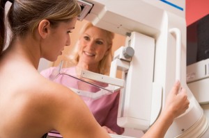
 The mammogram, or x-ray of the breast and surrounding tissue, is the most effective diagnostic tool for breast cancer that we have today. All women should receive annual mammograms beginning at age 40, or earlier with a family history of breast cancer.
According to www.breastcancer.org, mammograms have been shown to lower the risk of death from breast cancer by 35% in women over age 50. It also means that more women who are found to have breast cancer early can keep their breasts.
In 2009, the U.S. Preventive Services Task Force questioned the need for mammograms in women under 50, and they recommended that screening mammograms begin at 50 instead of 40. Several prominent groups, including the American Medical Association and the American College of Obstetricians and Gynecologists, have emphatically stated that screening needs to begin at 40 instead of 50.
One risk of mammography is the rate of false negatives and false positives among younger women or women with dense breast tissue. Dense breasts can hide cancers, and mammograms can identify a perfectly normal variation in breast tissue and raise the alarm that it’s cancerous. Because of these mammogram drawbacks, we recommend that you not only perform monthly self-exams, but you should also have a secondary form of breast screening done, such as an ultrasound or an MRI.
Many women worry about the pain, but for most women it is merely uncomfortable for a few minutes. A mammogram compresses your breast (to reduce the thickness of the tissue) between two x-ray plates that are attached to a camera that takes photos of your breast. More than a couple pictures may be necessary for younger women or those with dense breasts.
From beginning to end, a mammogram takes about 20 minutes and involves much less radiation exposure today than in years past. According to the American Cancer Society, the radiation received during mammogram is about the same amount a person naturally gets in a 3-month period.
Typically, at least one radiologist reads your mammogram, and if two read it, the chances of missing a problem go down. If you are concerned, you can also have your mammogram analyzed by a computer through computer-aided detection (CAD). Software reviews mammogram images and marks areas of suspicion, and then the radiologist examines each area to see if it needs further evaluation.
Have you had a mammogram, and if so, do you have any words of advice?


