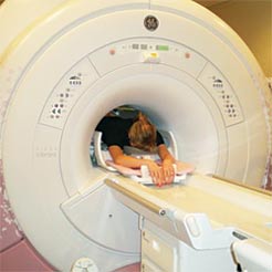
 Unlike a mammogram, which uses x-rays to create images of the breast, breast MRI uses magnets and radio waves to produce detailed 3-dimensional images of the breast tissue. Before the test, you may need to have a contrast solution (dye) injected into your arm through an intravenous line. The solution will help any potentially cancerous breast tissue show up more clearly.
Cancers need to increase their blood supply in order to grow. On a breast MRI, the contrast tends to become more concentrated in areas of cancer growth, showing up as white areas on an otherwise dark background. This helps the radiologist determine which areas could possibly be cancerous. More tests may be needed after breast MRI to confirm whether or not any suspicious areas are actually cancer.
For the breast MRI, you lie on your stomach on a padded platform with cushioned openings for your breasts. Each opening is surrounded by a breast coil, which is a signal receiver that works with the MRI unit to create the images. The platform then slides into the center of the tube-shaped MRI machine. You won’t feel the magnetic field and radio waves around you, but you will hear a loud thumping sound. You will need to be very still during the test, which takes around 30 to 45 minutes.
Because the technology uses strong magnets, it is essential that you remove anything metal — jewelry, snaps, belts, earrings, zippers, etc. — before the test. The technologist also will ask you if you have any metal implanted in your body, such as a pacemaker or artificial joint.
Where to have breast MRI?
It’s important to have breast MRI done at a facility with:
- MRI equipment designed specifically for imaging the breasts. Not all imaging centers have this; instead, many have MRIs used for scanning the head, chest, or abdomen.
- The ability to perform MRI-guided breast biopsy. If the breast MRI reveals an abnormality, you’ll want to have an MRI-guided breast biopsy (a procedure to remove any suspicious tissue for examination) right away. Otherwise, you’ll need to have a breast MRI again at another facility that offers an immediate MRI-guided breast biopsy.
See the MRI at The Charleston Breast Center Below


I was wondering if a MRI is still a valid diagnostic tool when scanning a sgap reconstructed breast? Also wondering the same thing about reconstruction using only fat grafting. Does this affect the MRI image and the ability to read it?